Pelvic Venous Disorder (PeVD) / Pelvic congestion syndrome Diagnosis
The diagnosis of Pelvic Venous Disorder (PeVD) or Pelvic Congestion Syndrome (PCS) needs a stepwise approach from the index PCS symptom to the effect on quality of life. The anatomical compressions seen in the veins carrying blood from the legs, ovaries, left kidney and pelvis are seen normally in many symptom-free women on CT and MRI scans. Enlarged ovarian and pelvic veins are also often seen on CT and MRI scans in women who have had children. It is the functional effect on the blood flow within the veins in the pelvis that must be identified and measured, along with the quality of pelvic floor function that may influence pooling of vein blood. This must then be correlated with the specific symptoms a patient has to accurately diagnose the type of pelvic vein disorder a patient is suffering from.
The tests we offer are tailored for the diagnosis of various aspects of the condition:
- Trans-abdominal -trans vaginal duplex ultrasound or PCS scan
- Dynamic Pelvic floor ultrasound scan
- Venous incompetency duplex ultrasound scan
- Tilt-table Test
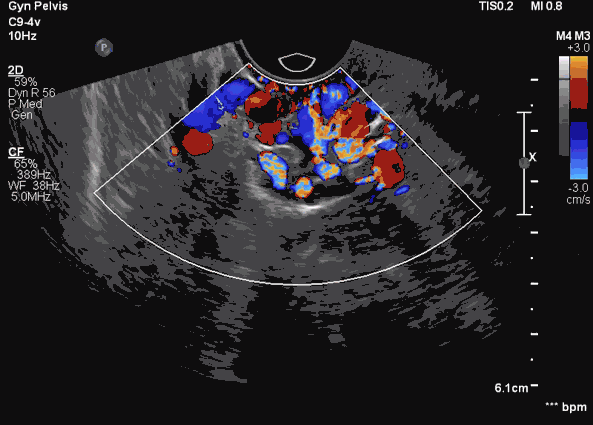
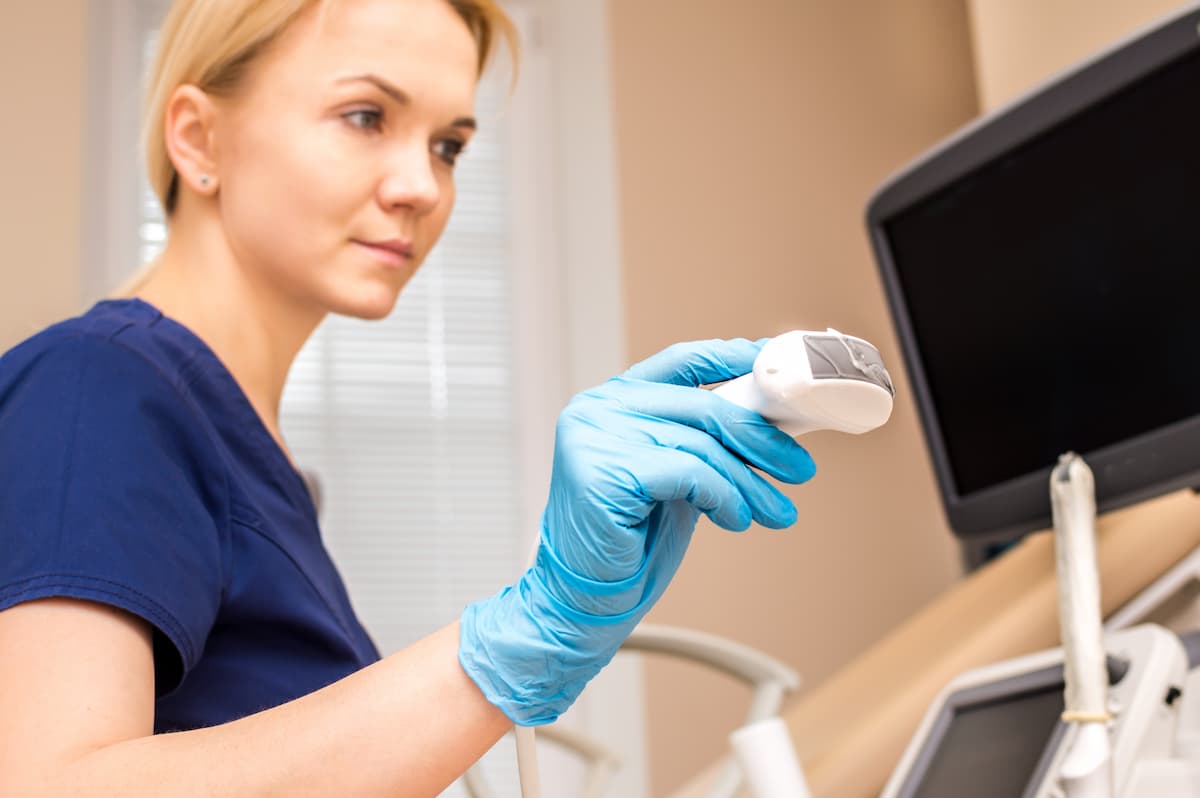
Trans-abdominal -trans-vaginal duplex ultrasound
The Trans-abdominal-transvaginal duplex ultrasound scan, which identifies abnormal blood flow in the veins of the ovary, kidney, and pelvis along with diseases in the pelvic organs that may mimic Pelvic Venous Disorder (PeVD) or Pelvic Congestion Syndrome (PCS), is considered by specialists to be the current gold-standard for making a diagnosis. Our team at the Venus Clinic have been international trainers for this type of ultrasound scanning for PCS in the UK and in Asia and follow a structured well practised protocol to make the diagnosis.
Dynamic pelvic floor ultrasound scan
We also offer assessments of the pelvic floor muscles and organs with dynamic pelvic floor ultrasound scanning and hope to offer targeted ultrasound duplex scans for Pelvic venous leaks soon as well. These allow us to assess the action of the pelvic floor muscles and identify specific sites of leakage of venous blood from the pelvis to the groin and legs.
Venous incompetency duplex ultrasound scan
Some patients with Pelvic Venous Disorder (PeVD) or Pelvic Congestion Syndrome (PCS) have pelvic veins leaking blood into the back of the legs and groin. These are usually visible as varicose veins. The venous incompetency duplex ultrasound scan maps these diseased veins to help plan the treatments needed.
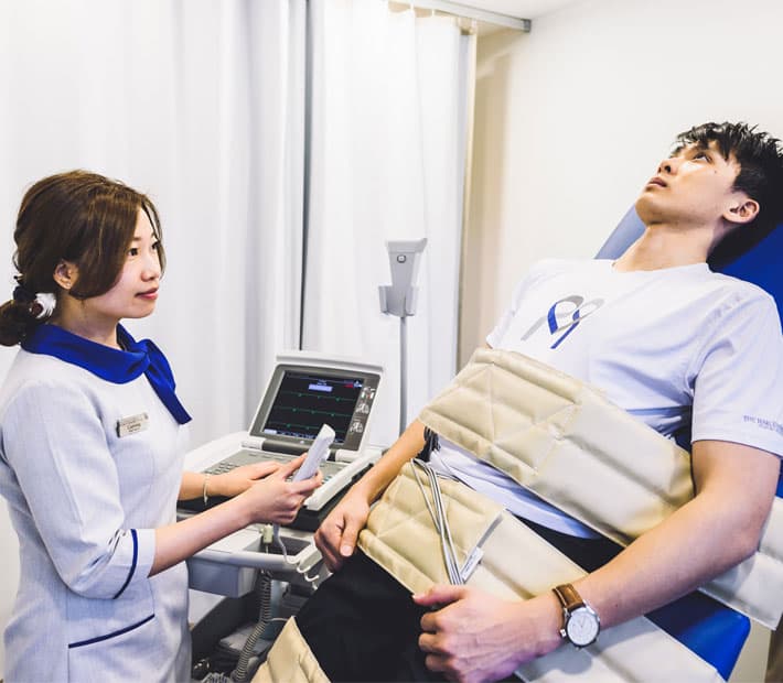
Tilt-table Test
Postural Orthostatic Tachycardia Syndrome (POTS) is a condition that affects blood flow. POTS causes the development of symptoms such as but not limited to light headedness, fainting and an uncomfortable, rapid increase in heartbeat. Many women with POTS also suffer from Pelvic Venous Disorder (PeVD) or Pelvic Congestion Syndrome (PCS), and report symptoms like brain fog, easy fatigue, palpitations, abdominal bloating, swelling of the legs, excessive or no sweating and rarely heart rhythm problems.